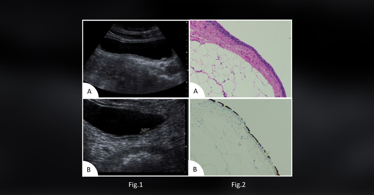
Case presentation A 66-year-old man was presented to the hospital for an incidental bladder lesion. The lesion was found during a trans-abdominal ultrasound examination, which was performed to investigate renal impairment. The patient denied any symptoms of previous urinary tract infections or lower urinary tract symptoms (LUTS).
Previous medical history Chronic kidney disease Congestive heart failure Type 2 diabetes mellitus Atrial myxoma observed several years back Social history Smoker Clinical examination Urine microscopy : The examination results showed microscopic hematuria and a few atypical urothelial cells. Ultrasound imaging of urinary bladder : Imaging revealed an echo-dense lesion on the posterior wall of the bladder, measuring 4.9 × 6.9 mm in size (Figure 1). Cystoscopy with bilateral retrograde pyelography Excluded abnormality of renal tract Showed normal bilateral…