
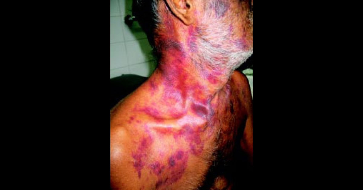
These are images of a 60 years old male presented with purpuras, petechiae, ecchymotic patches over the face, chest, shoulders and back since one and half years along with a history of swelling of limbs and breathlessness. Clinical examination It revealed ECOG performance status of 3, skin lesions, hepatomegaly and pedal oedema. Investigations showed grade 1 anaemia, normal renal and liver functions tests. Bone marrow aspirate was suggestive of 18% plasma cells.
Skeletal survey revealed multiple lytic lesions in skull and mandible. Protein electrophoresis (SPE) showed sharp and dense M band (2.1g/dl) in serum and narrow M band in urine. Abdominal fat biopsy showed normal blood vessels with no amyloid deposits. Skin biopsy showed amyloid deposits in dermis and vessels which showed apple green birefringence on Congo Red stain under polarized light features compatible with amyloidosis.…