
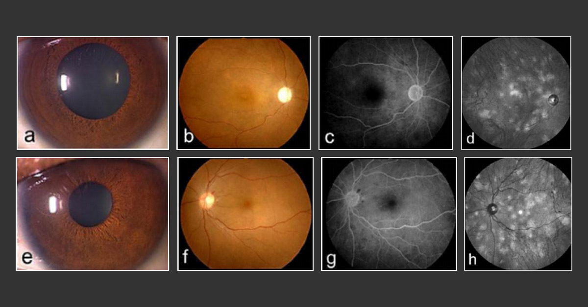
A 42-year-old woman was referred to the hospital due to complaints of transient blindness in her left eye. At the age of 33, the patient had a history of Systemic Lupus Erythematosus ( SLE ) and central retinal artery occlusion in her right eye.
Initial ophthalmic examination Best-corrected visual acuity of 1.2 in the left eye and no light perception in the right eye Normal ocular pressure and anterior segments Fundus examination showed no specific retinochoroidal abnormalities except for optic disc atrophy due to central retinal artery occlusion in her right eye and a peripapillary small hemorrhage in her left eye Fluorescein angiography revealed no dye leakage and pooling Near-infrared reflectance (NIR) showed multiple bright patchy lesions in the choroid of the posterior pole and in the midperiphery of the fundus in both eyes (fig. 1d, h) OCT Imaging of lesions revealed irregular,…





