
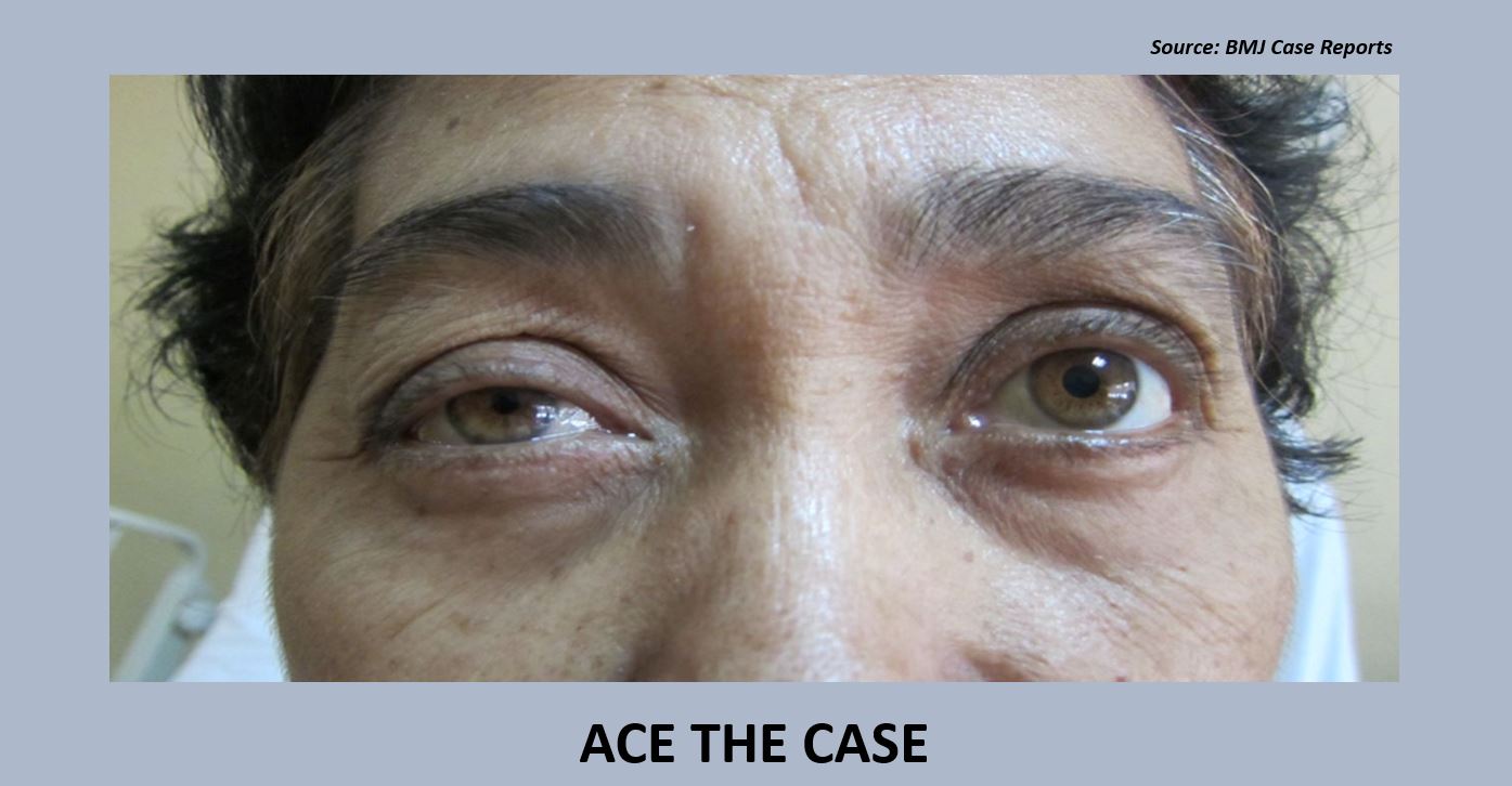
Case presentation A 63-year-old woman reported to the hospital with a mass in the right anterior portion of the neck that had increased in size rapidly over the past two months. She also reported having dysphagia without any associated dyspnea, hoarseness, or thyrotoxic symptoms. Pre-operative CT scan A well-defined mass (7.6×8.8×7.3 cm) was revealed, which enhanced the coarse calcifications seen on the right thyroid lobe, encircled the right common carotid artery, infiltrated the brachiocephalic, right internal jugular, and external jugular veins, and extended up to the thoracic inlet. Multiple enlarged lymph nodes were observed in the neck.
Lung metastases were observed. Biopsy Fine-needle aspiration biopsy of the thyroid gland was suggestive of papillary thyroid carcinoma. However, the nature and extent of the metastasis were suggestive of anaplastic carcinoma. A diagnosis of…





