
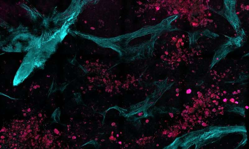
Multiphoton Microscopy works by delivering laser light to tissue. Though the laser has high peak intensity, it is delivered in very short pulses in order to keep the average power small and not cause tissue damage. As different tissue components interact with the laser light, they emit signals that are then retrieved by the microscope to create an image.
Once the images are acquired, automated image processing algorithms can be used to reveal unique textural features. These features, which are not visible in the images acquired with standard operative imaging tools, can be analyzed with statistical models to classify the tissue as healthy or diseased. A key strength of the approach is that the image acquisition and analysis are based on components of the tissue itself—such as cells or collagen, a protein that forms connective tissue—rather than on contrast dyes that have been added to…





