
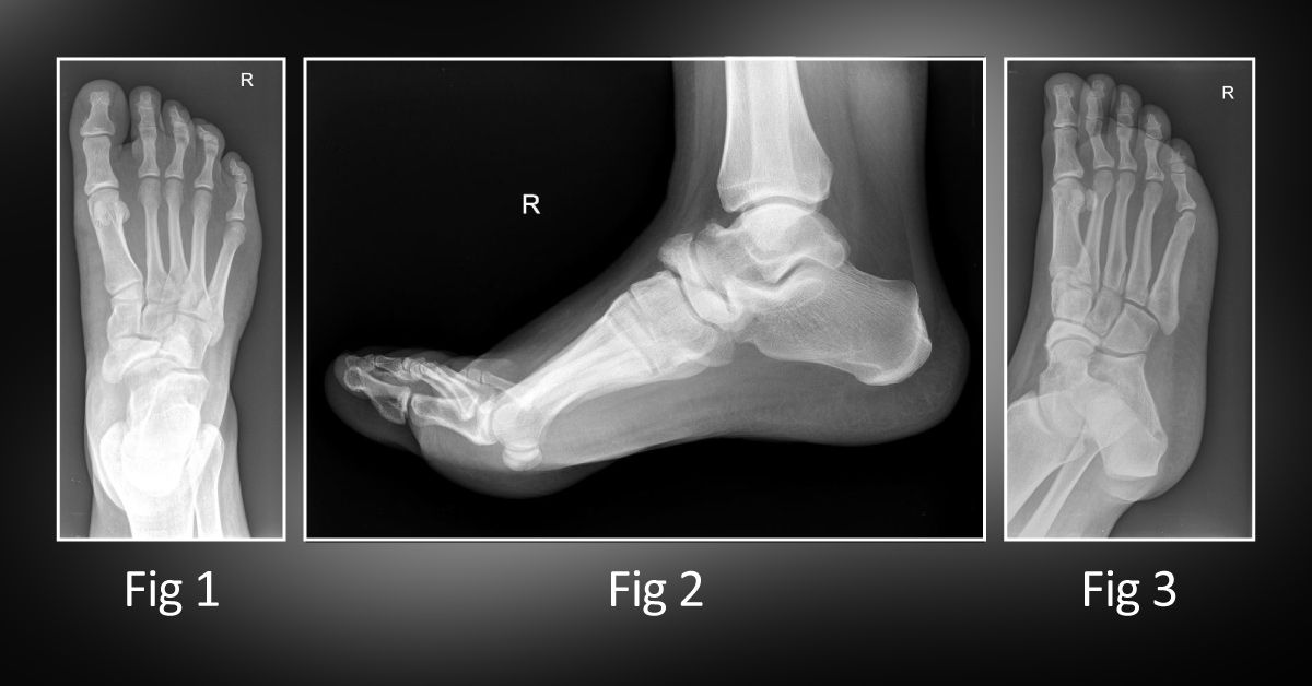
A 30-year-old male presented to an orthopedics OPD with a history of fall with sprain injury. He was advised an X-ray of his right foot. The X-ray revealed patchy sclerosis at the navicular bone . The frontal (Fig. 1), lateral (Fig. 2), and oblique (Fig.
3) X-rays are attached for your reference. ##Disclaimer## The content provided on Docplexus is intended for educational purposes only. While we strive to ensure its accuracy, we make no warranties, explicit or implied, about the suitability, reliability, or adequacy of the information, including data visualization elements and graphics. We do not endorse any medical treatment, product, ideas or service, including the content from our industry partners. We do not associate with the views of any individual, community, organization, industry partner or political party. Unauthorized use, reproduction, or distribution of the content outside…





