
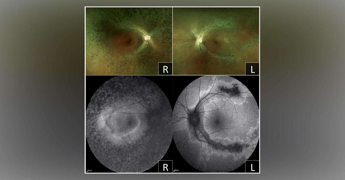
A 24-year-old female presented with a complaint of a narrowed visual field for the right eye since she was a teenager. Her left eye was normal. The systemic, ocular, medical, or familial history was unremarkable. At the first visit, the best-corrected visual alertness in the decimal fraction was 1.2 in both eyes with a r efraction of -0.75 OD and -3.75 OS , respectively.
Fundoscopic examination along with fundus autofluorescence (FAF) imaging technique showed different abnormalities in both eyes. (Cover image) Left eye: Peripapillary retinochoroidal atrophy extending along the retinal vein, bone spicule pigment accumulation distributed at distal parts of the atrophic areas, increased autofluorescence surrounding the region of RPE atrophy; macula and optic disc were intact. Right eye: Unaffected macula surrounded by peripheral retinal pigmented epithelium (RPE) atrophy with diffuse…





