
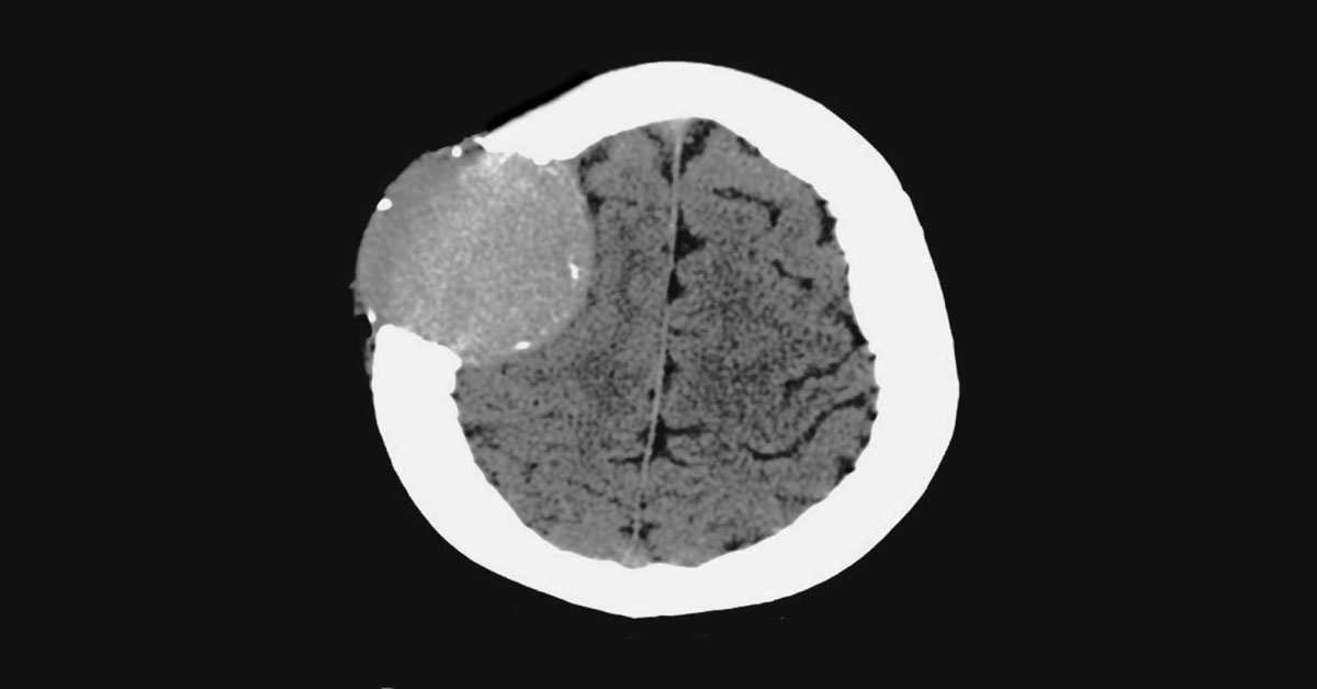
A 42-year-old woman presented to a neurosurgeon with a painless, coin-sized lump on her scalp. The lump was initially noticed by the patient’s hairdresser four months prior to the clinical evaluation.
Physical Examination A firm, nontender, subcutaneous nodule with a diameter of 6 cm Absence of any neurologic deficits Other Diagnostic Tests Analysis of CBC, complete metabolic profile, and urine: normal Computed Tomographic (CT) Analysis A CT scan of the lump revealed the following results: Destructive, well-demarcated, soft-tissue mass of 5 cm by 4 cm The mass had peripheral bony fragments emerging from the right frontal calvarium No evidence of a midline shift Surgical Procedure The mass was further excised, and a cranial mesh was placed over the calvarial deficit Histologic examination Histological analysis of the lump revealed 20% plasma cells Skeletal survey Multiple lytic lesions…





