
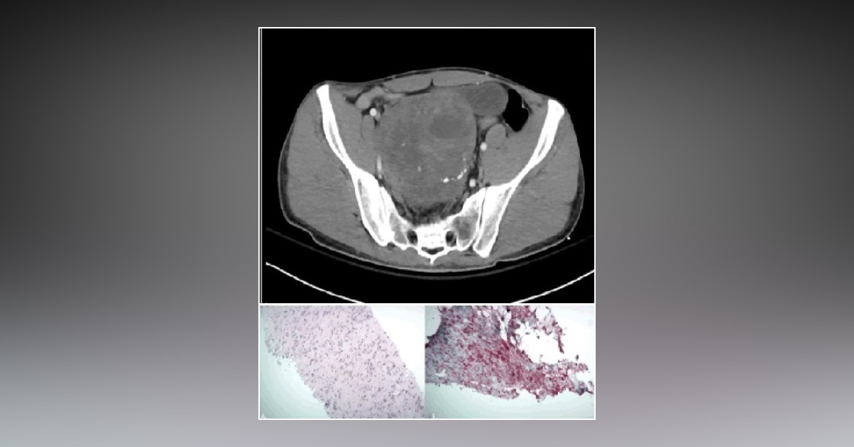
A 28-year-old male presented to the trauma care center after a motor vehicle collision. Subsequent CT scan revealed a fractured c1 vertebrae. However, during abdominal CT scanning, a solid mass of 11 cm × 9.5 cm × 12.6 cm was observed, which had areas of calcification and hemorrhage. CT scan image has been provided. There were no symptoms of polyuria or dysuria. The patient also denied any sensory or motor deficits in lower extremities. A CT-guided biopsy was performed.
Pathological evaluation revealed S-100 positive and desmin/vimentin negative spindle cell lesion (image provided). Bilateral stenting had been placed by urologists at the beginning of treatment. Exploratory laparotomy was performed, which utilized the lower midline abdominal incision. The densely adherent mass was removed from the pelvis intact. Pathological staining and sectioning images have been provided. Samples were…





